How Do Arteries and Veins Work? Blood Vessel Structure and Function
Learn More About Simplified Science PublishingBlood vessels are muscular and flexible tubes that create your blood transport system and is made of arteries, capillaries, and veins.
Cardiovascular System Review
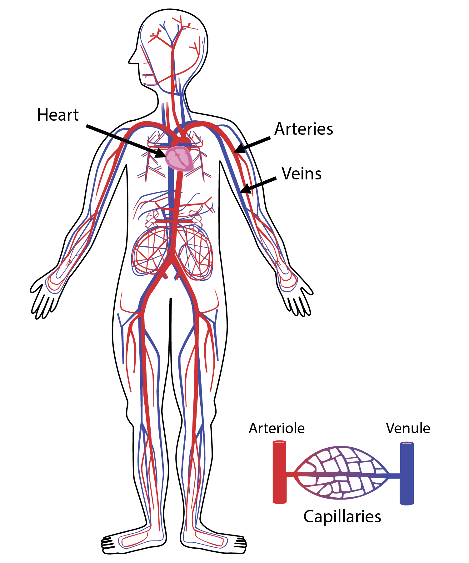
In Greek, the word “kardia” means heart and in Latin, the word “vas” means vessel. This Greek and Latin combination creates a straight-forward translation for the cardiovascular system: the heart and blood vessels. These organs create your blood transportation system and tirelessly move five to six liters of blood on a full tour of your body nearly every minute.
In Greek, the word “kardia” means heart and in Latin, the word “vas” means vessel. This Greek and Latin combination creates a straight-forward translation for the cardiovascular system: the heart and blood vessels. These organs create your blood transportation system and tirelessly move five to six liters of blood on a full tour of your body nearly every minute.
This article will focus on the structure and function of blood vessels and you can view this article to read more about heart anatomy.

Artery Structure and Function
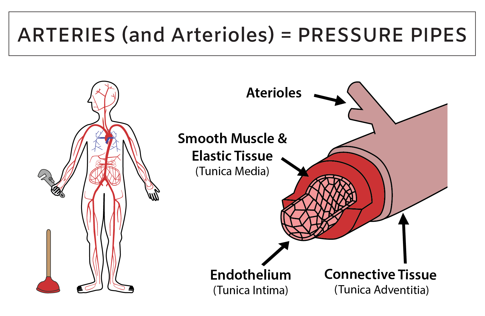
Within the walls of every modern building is an organized system of pipes that supply pressurized water to the sinks, toilets, and showers. Comparatively, your internal plumbing is chaos. Every square inch of your body contains a dense tangle of blood vessel pipes. The pipes that supply newly oxygenated blood to all of your tissues are called arteries. Arteries support the life of every cell in your body and problems with your arterial plumbing, such as clogs or cracks, can dramatically impact your health.
The defining characteristic of arteries is that they carry blood away from the heart. Therefore, arteries are your pressure pipes that handle large changes in blood pressure with each heartbeat. During the pump (systole), the heart’s ventricles send blood shooting into the arteries, which briefly increases pressure. During the refill (diastole), the heart muscles are relaxed and pressure in the arteries is decreased. This cycle of pressure changes stretches and relaxes the artery walls, thereby creating your pulse and blood pressure. When you press your fingers against your wrist or neck, the pulse that you feel pressing against your hand is due to the repeated surging and slowing of blood within your arteries.
Almost every artery carries newly oxygenated blood to resupply the body’s tissues, but there are two exceptions: the pulmonary arteries, which carry deoxygenated blood from the heart to the lungs and umbilical arteries, which carry deoxygenated blood from a pregnant woman’s fetus to the placenta.

Artery walls are made of three main layers of cells that are called tunics. The word tunic means a close-fitting coat, so you can think of the blood vessel layers as your cardiovascular system’s clothing layers that change in thickness depending on their location and environment.
Every artery and arteriole has three tunics: intima, media and adventitia. Tunica intima is the inner-most layer and it is made of a single layer of endothelial cells. This layer is similar among all arteries and functions as the base layer for each blood vessel outfit. The tunica media is the middle layer and it is made of smooth muscle and elastic tissue. The thickness of this layer changes dramatically depending on an artery’s location. Arteries that are near the heart receive strong bursts of blood with each heartbeat and they have a thick smooth muscle layer and a strong elastic layer that provide strength and flexibility to handle the high blood pressure. The arterial pipes that carry blood throughout the body branch to become narrow tubes called arterioles.
In contrast, arteries and arterioles that are far away from the heart are not exposed to high blood pressure and have a thin middle muscle and elastic layer. Finally, the tunica adventitia is the outer coat and it is made of tough connective tissue. This layer is slightly thicker in major arteries near the heart and very thin in arterioles.
Capillaries Structure and Function
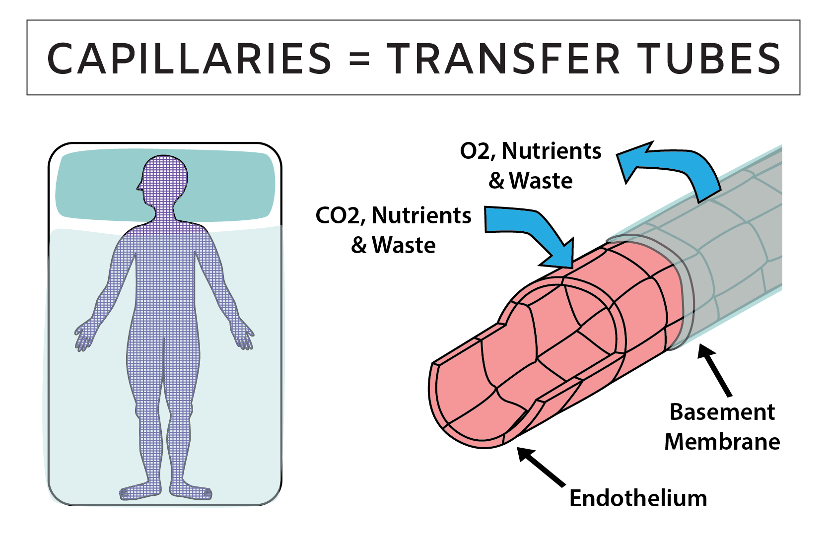
In order to survive, every living cell in your body needs to be located near a network of thin blood vessel tubes that are called a capillary beds. These so-called beds are more like tiny tissue hammocks that cradle the cells throughout your body. How tiny? The width of a capillary is less than the width of the thinnest human hair. Look at the hair on your arm—your organs are enmeshed in capillary beds whose threads are thinner than those hairs.
In many ways, capillaries connect your internal tissue to the outside world. Arteries and veins are the major highways that carry blood throughout the body, but your capillaries allow water, nutrients, hormones and waste to transfer between the bloodstream and your body’s cells. Therefore, capillaries are your transfer tubes and their function is essential for every organ. Without capillaries, the air you breathe, the water you drink and the food that you eat would have no way of reaching the cells that depend on them to function.

Vein Structure and Function
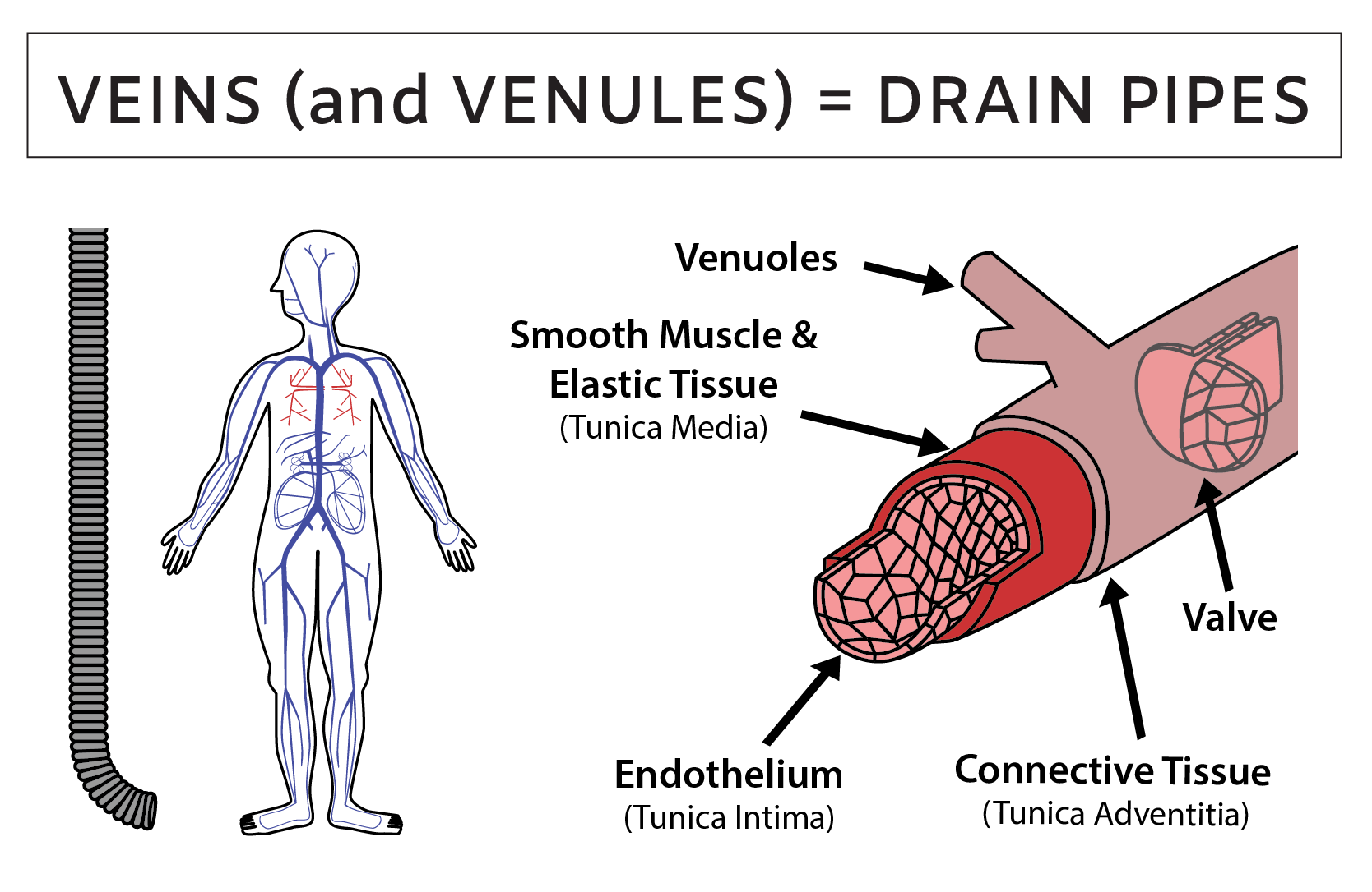
Once blood has squeezed through the capillaries, it flows into the drain pipes of the circulatory system which are called the veins and venules. Modern water drainage systems receive liquid from sinks, showers, toilets and soil, send it through filters to remove toxins and waste, and then send it back into the main water supply. Veins function similar to a drainage system because they take blood that has been filtered through the body’s tissues, send it through additional organ filtration systems to remove toxins and waste and then send the blood back to the heart to begin the journey again.

Related Content:
- What is Blood Made Of? Review Blood Components and Functions
- How Does the Heart Work? Review Heart Structure and Function
- What is a Heart Attack? Heart Attack Causes and Prevention Tips
- Heart Electrical System: ECG and Sequence of Electrical Conduction
- What is Aerobic Respiration and Why is it Important?
- How Does the Diaphragm Work? Diaphragm Structure and Function
References:
- Harrison's Principles of Internal Medicine, 18th Edition. Longo DL, Fauci AS, Kasper DL, Hauser SL, Jameson J, Loscalzo J. eds.
- Anatomy, Physiology, and Disease: An Interactive Journey for Health Professions , 2nd Edition. Bruce J. Colbert, University of Pittsburgh, Johnstown.
Create professional science figures with illustration services or use the online courses and templates to quickly learn how to make your own designs.
Interested in free design templates and training?
Explore scientific illustration templates and courses by creating a Simplified Science Publishing Log In. Whether you are new to data visualization design or have some experience, these resources will improve your ability to use both basic and advanced design tools.
Interested in reading more articles on scientific design? Learn more below:
Content is protected by Copyright license. Website visitors are welcome to share images and articles, however they must include the Simplified Science Publishing URL source link when shared. Thank you!


