

How Do Lungs Work? Lung Structure and Function
Learn More About Simplified Science PublishingThe lungs are a spongy network of air tubes and blood vessels and the three main lung structures that allow for gas exchange are bronchi, bronchioles, and alveoli.
Evolution of lungs
When creatures first wriggled out of the sea and onto dry land, they were gifted with a crucial adaptation: primitive lungs. These lungs were not the efficient air bags that we have today. They were a loose collection of blood vessels that were originally created to absorb tiny bits of oxygen from water running across them.
When early amphibious creatures began to spend more time on land, the crude network of blood vessels and tubes eventually evolved into a tightly woven mesh that more closely resembled the lung structures that allow humans to absorb oxygen from the air instead of water.
Lung structure review
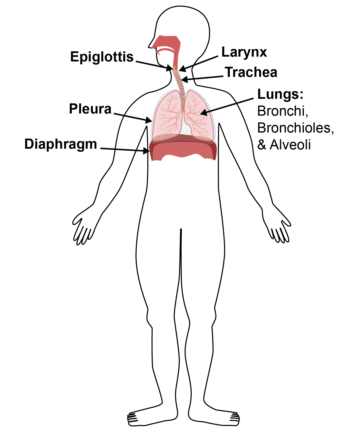
The lungs are a spongy network of air tubes and blood vessels and the three main lung structures that allow for gas exchange are bronchi, bronchioles and alveoli. The primary bronchi are similar to two penne noodle tubes that split off from the central airway and they shuttle air into the left and right lungs. These bronchi tubes then split off into increasingly thinner spaghetti-width tubes, which are called your bronchioles. Lastly, the bronchioles guide air into millions of microscopic sacs called alveoli, which are your gas exchange meatballs.

How do lungs work?
When you take a breath, air moves past the main tube (called the trachea) and branches into the two smaller bronchi tubes. The brochi shuttle air into the right and left lung lobes and from there, the air is sent into the network of tiny bronchiole tubes that resemble slender noodles. As you inhale, your chest and tiny tubes have expanded to compensate for the increased pressure and the air is forced to move into tiny sacks called alveoli. The alveoli are the microscopic meatballs in this somewhat silly pasta analogy and take on the most important role for your entire respiratory system: the exchange of oxygen and carbon dioxide.
One key feature of the alveoli is that they are packed with tiny air tubes and also enmeshed in a dense tangle of thin blood vessels. The thinnest blood vessels surround each alveoli air sac cluster and as the blood rushes past the alveoli air tubes, it release the excess carbon dioxide and absorbs oxygen molecules before being pumped back into your heart to be distributed to the rest of your body.
Related Content:
- How Does the Diaphragm Work? Diaphragm Structure and Function
- What are Pleura? Lung Pleura Structure and Function
- What is the Epiglottis? Epiglottis Structure and Function
- How Does the Heart Work? Review Heart Structure and Function
- What is Blood Made Of? Review Blood Components and Functions
- What is Aerobic Respiration and Why is it Important?
References:
- Harrison's Principles of Internal Medicine, 18th Edition. Longo DL, Fauci AS, Kasper DL, Hauser SL, Jameson J, Loscalzo J. eds.
- Anatomy, Physiology, and Disease: An Interactive Journey for Health Professions , 2nd Edition. Bruce J. Colbert, University of Pittsburgh, Johnstown.
Create professional science figures with illustration services or use the online courses and templates to quickly learn how to make your own designs.
Interested in free design templates and training?
Explore scientific illustration templates and courses by creating a Simplified Science Publishing Log In. Whether you are new to data visualization design or have some experience, these resources will improve your ability to use both basic and advanced design tools.
Interested in reading more articles on scientific design? Learn more below:


Content is protected by Copyright license. Website visitors are welcome to share images and articles, however they must include the Simplified Science Publishing URL source link when shared. Thank you!